lv filling pressure Left ventricular filling pressure is the pressure that fills the ventricle in diastole and determines stroke volume according to the Frank-Starling mechanism. In patients with HF, . See more +371 67095405 +371 67095689 pasts [at] fm.gov.lv Smilsu street 1, Riga, LV-1919. E-address. Read more; Got a question? To send a question, fill in all mandatory fields in the question form! State authority in accordance with the Application Law has the right not to answer the received letter (application) if the sender has not indicated the .
0 · raised lv filling pressures
1 · lv filling pressure normal range
2 · left ventricular filling pressure chart
3 · increased lv filling pressure
4 · increased left ventricular filling pressure
5 · elevated lv filling pressures
6 · elevated left ventricular filling pressures
7 · elevated left sided filling pressures
+371 67095405 +371 67095689 pasts [at] fm.gov.lv Smilsu street 1, Riga, LV-1919. E-address. Read more; Got a question? To send a question, fill in all mandatory fields in the question form! State authority in accordance with the Application Law has the right not to answer the received letter (application) if the sender has not indicated the .
Left ventricular diastolic dysfunction (LVDD) plays a key role in the pathophysiology of heart failure (HF). It is caused by impaired left ventricular (LV) relaxation with or without reduced restoring forces and increased LV chamber stiffness leading to the inability of the ventricle to fill adequately . See more

shop dolce and gabbana
Left ventricular filling pressure is the pressure that fills the ventricle in diastole and determines stroke volume according to the Frank-Starling mechanism. In patients with HF, . See moreIdentification of elevated LVFP at rest or during exercise is pivotal for the diagnosis of HFpEF, which gained additional interest since medical . See moreA number of echocardiographic parameters may be used to differentiate between normal and elevated LVFP. All the recommended . See more

raised lv filling pressures
Figure 1 shows the algorithm recommended by the European Association of Cardiovascular Imaging (EACVI) to evaluate LVFP.2 Importantly, the recommendations advocate careful consideration of all available clinical, 2D, and Doppler data to . See moreNoninvasive Assessment of LV Filling Pressure. Elevated left ventricular (LV) filling pressure may be used to confirm a diagnosis of heart failure. We .
For normal cardiac performance, the left ventricle (LV) must be able to eject an adequate stroke volume at arterial pressure (systolic function) and fill without requiring an elevated left atrial (LA) pressure (diastolic function). .Results: Mean left ventricular ejection fraction (LVEF) was 47%, with 209 patients having an LVEF <50%. Invasive measurements showed elevated LV filling pressure in 58% of patients. Invasively measured pressure (pulmonary artery wedge pressure [PCWP] or pre-A-wave LV end-diastolic pressure >12 mm Hg) was used as the gold standard. Results: Mean . Echocardiography is widely used to evaluate left ventricular (LV) diastolic function in patients suspected of heart failure. For patients in sinus rhythm, a combination of several .
lv filling pressure normal range
left ventricular filling pressure chart
The term left ventricular filling pressure (LVFP) refers to the LV pressures during diastole. They are illustrated in Fig. 13.3 and include LV minimal pressure, pre-A wave pressure, and LV end .

Figure 1. LA and LV Pressures. The lowest left ventricular (LV) pressure is the minimum pressure (LV min). After mitral valve opening, LV pressure rises, producing rapid filling wave (RFW) to LV pre–A-wave pressure (LV .
Left atrial volume index, in combination with flow velocities and tissue Doppler velocities, was used to estimate LV filling pressure. Invasively measured pressure was used .
Left ventricular (LV) diastolic dysfunction is a condition of impaired LV relaxation and increased LV chamber stiffness, which can lead to elevated LV filling pressures. This topic . Predicted probability of PCWP > 15 mmHg according to the developed scoring system in the whole population. Scoring points are represented on the x-axis; probability of ILFP is represented on the y-axis. .Left ventricular (LV) diastolic function is characterized by LV relaxation, chamber stiffness, and early diastolic recoil, all of which determine LV filling pressure. Echocardiographic signals significantly associated with LV .After the ventricle’s compliance decreases for an extended length in time, with increasing the LV filling pressures and preload, the LAP will increase to ensure sufficient blood is being filled in the LV. The LAP increase allows the rapid .
(B) In the algorithm to estimate LV filling pressure, we incorporated recommendations from recent guidelines (5), setting criteria of elevated LV filling pressures as either E/A ratio ≥2 in the presence of myocardial disease, or if E/A is <2, at least 2 of the 3 parameters shown must be above cutoff values (table in A). The algorithm was . Estimation of left ventricular filling pressure in patients with mitral valve disease and mitral annular calcification. In patients with isolated mitral stenosis, LVEDP is usually normal but LA pressure is elevated. IVRT which corresponds to the time interval between the aortic component of the second heart sound and the opening snap of the .
The following are key points to remember about pulmonary arterial wedge pressure and left ventricular end-diastolic pressure for assessment of left-sided filling pressures: The terms “pulmonary arterial wedge pressure” (PAWP) and “left ventricular end-diastolic pressure” (LVEDP) are often used interchangeably to describe left-sided . Background The utility of Doppler velocities across the patent foramen ovale (PFO) to estimate left ventricular (LV) filling pressure is not well known. Methods The best cut-off value of peak interatrial septal velocity across a transeptal puncture site measured by transesophageal echocardiography for estimating high mean left atrial (LA) pressure (≥ 15 .
Invasive methods of measuring LV relaxation and filling pressures are considered the gold-standard for investigating diastolic function. However, the high temporal resolution of trans-thoracic echocardiography (TTE) with widely validated and reproducible measures available at the patient’s bedside and without the need for invasive procedures .ASSESSMENT OF LV FILLING PRESSURES AND DIASTOLIC DYSFUNCTION GRADE (For full recommendation refer to the Left Ventricular Diastolic Function Guideline p. 281) Diastolic Function Assessment in Patients with Normal vs Abnormal LVEF In patients with a normal LVEF, the initial assessment is to determine the presence or absence of diastolic . Left atrial volume index, in combination with flow velocities and tissue Doppler velocities, was used to estimate LV filling pressure. Invasively measured pressure was used as the gold standard. Results: Mean left ventricular ejection fraction (LVEF) was 47%, with 209 patients having an LVEF <50%. Invasive measurements showed elevated LV .
Left ventricular diastolic dysfunction (LVDD) is a condition that affects your heart’s ability to fill up with blood before sending the blood out into your circulation. Your heartbeat has two .
increased lv filling pressure
In addition, LV filling pressure was derived from a regression equation that had previously correlated the medial E-e′ ratio with LV filling pressure in patients without HCM. 4 With the use of this regression equation, the mean difference between the calculated LV filling pressure and measured LAP was −0.5±6.9 mm Hg (95% confidence limits . A shortened early deceleration time indicates an increased LV operating stiffness. It is a hallmark of restrictive filling pattern and denotes poor prognosis in patients with myocardial infarction, dilated cardiomyopathy, heart transplantation, hypertrophic cardiomyopathy, and restrictive cardiomyopathy. 4 Both pseudonormalized and restricted filling patterns indicate a . Results: Mean LV ejection fraction (LVEF) was 47%; LVEF was . 50% in 209 patients.. Invasive measurements showed elevated LV filling pressure in 58% of the patients. Echo/Doppler data acquisition was feasible in 419 patients (93.1%); testing the ASE/EAE/EACI recommendations used 320 patients (after further excluding patients with mitral regurgitation, . When atrial contraction contributes significantly to LV filling, 9, 10 LVEDP can increase without an increase in mean LV diastolic pressure , which happens during an early stage of diastolic dysfunction with delayed LV .
Background— The estimation of left ventricular (LV) filling pressure from the ratio of transmitral and annular velocities (E/e′) after exercise echocardiography may identify diastolic dysfunction in patients who complain .
III. Echocardiographic Assessment of LV Filling Pressures and Diastolic Dysfunction Grade 281 IV. Conclusions on Diastolic Function in the Clinical Report 288 V. Estimation of LV Filling Pressures in Specific Cardiovascular Diseases 288 A. Hypertrophic Cardiomyopathy 289 B. Restrictive Cardiomyopathy 289 C. Valvular Heart Disease 290Left ventricular filling pressures are increased, and this increase is transmitted back through the pulmonary microvascular bed. As pressure in that bed is increased, transcapillary fluid flux tends to increase, increasing the risk of edema discussed previously. Further retrograde pressure transmission elevates pulmonary artery pressure .Background Increase in left ventricular filling pressure (FP) and diastolic dysfunction are established consequences of progressive aortic stenosis (AS). However, the impact of elevated FP as detected by pretranscatheter aortic valve replacement (TAVR) echocardiogram on long-term outcomes after TAVR remains unclear. Objective To understand the impact of elevated .
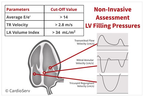
It occurs when your lower heart chambers don’t relax and fill with blood properly. . your ventricles don’t fill with blood as they should, and you may experience pressure buildup in your heart. This can progress to diastolic heart failure, resulting in fluid buildup in your lungs, abdomen and legs. . Left ventricular assist devices: .Left ventricular filling pressures are thereby increased. This increase will be transmitted back through the pulmonary microvascular bed. As pressure in that bed is raised, transcapillary fluid flux will tend to increase, raising the risk of edema (see earlier discussion). Further retrograde pressure transmission will elevate pulmonary artery .As LV filling pressure progressively elevate, the LAVI size will enlarge. Therefore, an enlarged LAVI is a marker of elevated LAP. In contrast, a small or normal LAVI is suggestive of normal LAP. The abnormal cut-off value for LAVI is > 34mL/m². Estimation of left ventricular filling pressure in patients with mitral valve disease and mitral annular calcification. In patients with isolated mitral stenosis, LVEDP is usually normal but LA pressure is elevated. IVRT which corresponds to the time interval between the aortic component of the second heart sound and the opening snap of the .
Diastolic function is a catch-all term referring to several different physiological processes that allow the left ventricle (LV) to fill with sufficient blood for the body’s current needs at a low enough pressure to prevent pulmonary congestion.Diastole (Table 1) actually begins in systole, as energy stored in titin within the myocyte and as torsion in the interstitial fibers of the . Backgrounds Assessment of left ventricular filling pressure (LVFP) is of clinical importance in patients with ST elevation myocardial infarction (STEMI). Although several echocardiographic parameters are recommended for the assessment of LVFP, validation of these parameters in patients with STEMI is missing. We aimed to investigate the clinical utility of .Markedly delayed LV relaxation in the setting of elevated LV filling pressures allows for ongoing LV filling in mid diastole and thus L velocity. Patients usually have bradycardia. When present in patients with known cardiac disease (e.g., LVH, HCM), it is specific for elevated LV filling pressures. However, its sensitivity is overall low.
While invasively measured filling pressure is the gold standard, echocardiographic measurement is a clinically accepted tool for estimating left ventricular filling pressure and has been adopted .
increased left ventricular filling pressure
Send flowers with same-day delivery to Las Vegas, NV & cities nationwide from Flowers by Coley, your local Las Vegas florist. 100% Satisfaction guaranteed. Get FREE local delivery with upgrade!
lv filling pressure|elevated left sided filling pressures


